Charles University, Faculty of Science, Prague, Czechia
Research
We integrate traditional helminthology and approaches of modern experimental biology. In the lab, we study host-helminth interactions from molecular, biochemical, or immunological perspectives. In the field, we focus on topics related to cercarial dermatitis (swimmer's itch).
Our main laboratory model species include:
● avian schistosomes (Trichobilharzia spp.)
● human schistosomes (Schistosoma mansoni)
● larval tapeworms (Taenia crassiceps, Mesocestoides corti)
> Avian schistosomes
BACKGROUND
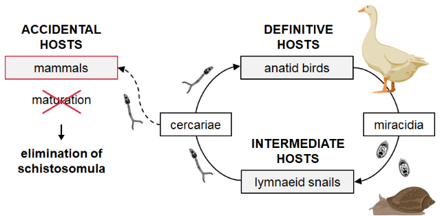
Avian schistosomes are worldwide distributed relatives of human blood flukes. The genus Trichobilharzia has become the most conspicuous, and it is recognized as the primary etiological agent of human cercarial dermatitis (CD), also known as swimmer’s itch.
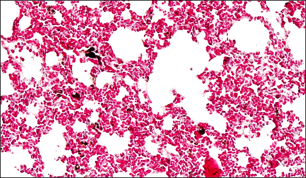
The life cycle of Trichobilharzia spp. includes two hosts: anatid birds and lymnaeid snails. The cercariae can also infect mammals, but they are soon eliminated as mammals represent unsuitable, accidental hosts. Nevertheless, they are valuable models for studying the host immune response against the parasites - either in the skin, lungs or central nervous system.
MODEL SPECIES
We maintain the life cycles of two Trichobilharzia species : the viscerotropic Trichobilharzia szidati (migrates through lungs to the intestinal wall) and the neurotropic Trichobilharzia regenti (migrates through the peripheral nerves and brain to the nasal mucosa).
Interestingly, Trichobilharzia regenti is the neuropathogenic species described by our team in 1998 from South Bohemia. It was named after a famous local brewery "Regent" founded in 1379. Adults of T. regenti live in the nasal mucosa (in the bill), where mating and egg laying occur. Thanks to its neurotropism exhibited also in mice, T. regenti has become a valuable model of helminth neuroinfections.

OUR FOCUS
We study different immunobiological aspects of Trichobilharzia infection in mice, such as:
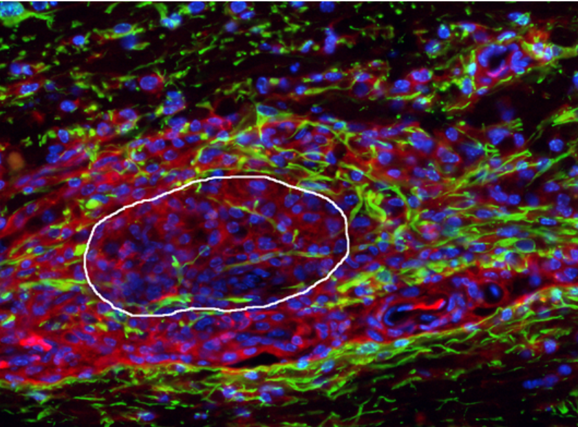
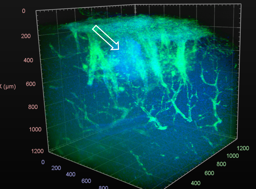
● the host immune response and neuropathogenicity of Trichobilharzia regenti in the central nervous system,
● the host immune response against Trichobilharzia cercariae penetrating the skin and the immunopathology associated with CD,
● the effect of Trichobilharzia infection on allergic and autoimmune diseases (asthma, experimental autoimmune encephalomyelitis),
● the role of Trichobilharzia-derived molecules, especially peptidases, in the aforementioned processes.


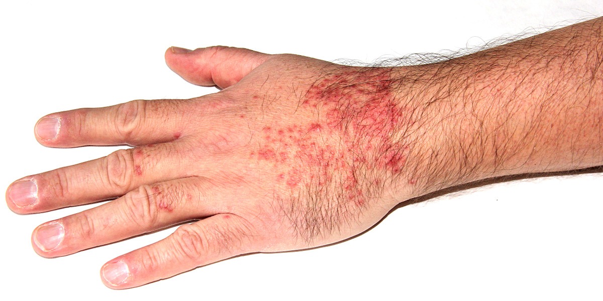
> Cercarial dermatitis
BACKGROUND
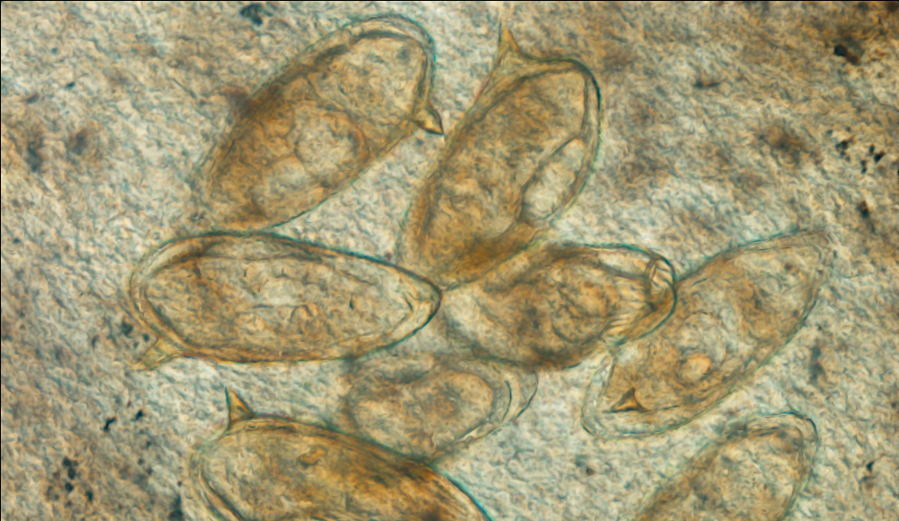
Cercarial dermatitis (CD; aka swimmer's itch) is a skin disease caused by schistosome larvae called cercariae. As they penetrate the mammalian skin, they trigger inflammation and allergic reactions, manifesting as unpleasant itching rash. Although the human species can trigger CD, avian schistosomes (mainly Trichobilharzia spp.) are the major causative agents. For more information, follow our comprehensive review or human clinical follow-up study.
OUR FOCUS
We study several field aspects related to CD, such as:
● diversity of avian schistosomes and their capacity to trigger CD,
● methods for environmental detection of avian schistosomes,
● monitoring of CD outbreaks in Czechia and managing affected sites.
> Human schistosomes
BACKGROUND
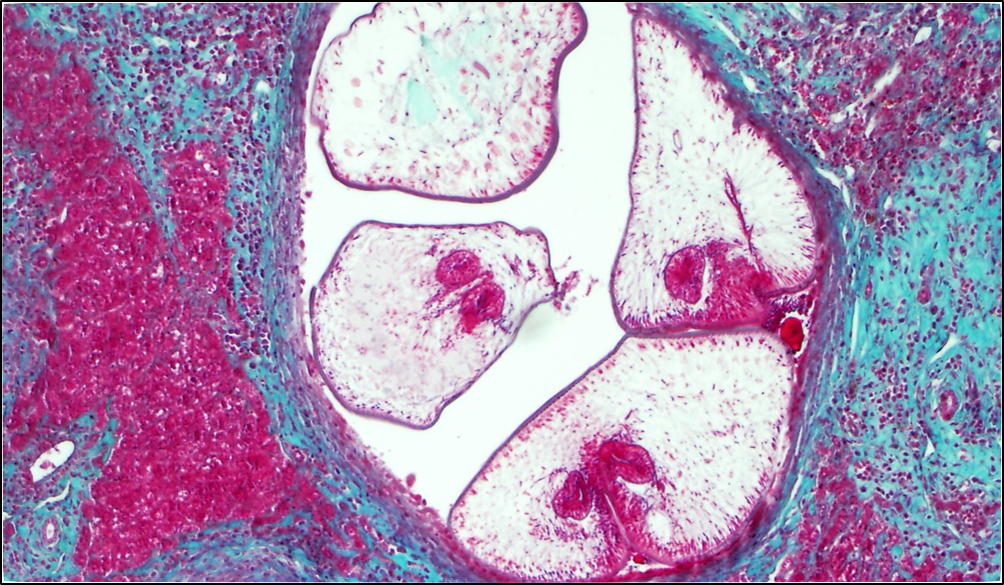
Human schistosomes affect millions of people, mostly in tropical areas. Schistosome eggs represent the major pathological agent as they are disseminated in the body and entrapped in the tissue. As for Schistosoma mansoni, the liver, and intestine often harbor large quantities of egg. Consequently, abdominal pain, diarrhea, and portal hypertension are usual symptoms.
OUR FOCUS
We have teamed up Laboratory of Molecular Parasitology (Czech University of Life Sciences, Prague) to explore:
● the egg-secreted proteins and identify novel bioactive molecules,
● differences among parasite eggs isolated from liver and intestine,
● pathology triggered by different S. mansoni geographical strains.
> Larval tapeworms vs. cancer
Currently, the research stream is suspended due to the absence of funding and (as a consequence) subteam members/students.
BACKGROUND
In recent years, research concerning the effect of various parasite species on cancer has experienced many developments, identifying infections and, subsequently, parasite-produced molecules, which can suppress the growth and metastasis of tumors in various direct or immunity-mediated ways across many types of cancer. Our research has revealed the capability of two tapeworm larvae infections to protect against an aggressive melanoma cell line in the mouse model.
MODEL SPECIES
Taenia crassiceps and Mesocestoides corti
These two cyclophyllidean tapeworms make for excellent laboratory models, as their larvae can asexually multiply in the peritoneal cavity of the experimental vertebrate host. They are prolific modulators of the host immune system due to the effect of their excretory-secretory products and other molecules/antigens being exposed. Besides that, T. crassiceps and its products have been shown to abrogate the spread of colon cancer and melanoma while also being used to ameliorate various autoimmune diseases, such as type 1 diabetes and multiple sclerosis.
OUR FOCUS
We study the effects of the tapeworm larvae infection and their excretory-secretory products on the growth and metastasis of various cancer cell lines, such as the B16F10 melanoma, ID8 ovarian carcinoma, and the Hepa 1-6 hepatocarcinoma, while also investigating the effects these products have on various immune cells, both in the mouse model and in vitro.
Address: Viničná 7, 12800 Prague 2, Czechia (see the map)
Lead contact: petr.horak@natur.cuni.cz